Small Animal Micro CT Imaging Experiment Service
Instruction manual
First Edition (Revised on April, 2016)
Information
Micro-CT (micro computed tomography) is a non-destructive 3D imaging technology. It can clearly understand the internal microstructure of the sample without destroying the sample. The biggest difference between ordinary clinical CT is the high resolution, the resolution can reach micron (μm). Micro-CT can be used to perform high-resolution (<10μm) X-ray imaging of bones, teeth, live small animals and various material devices without destroying the sample, obtain the detailed three-dimensional structure information inside the sample, thus showing the three-dimensional image of each part. The resolution is much higher than the clinical CT.
Service Content
√Research of animal osteoporosis (including changes in microstructure of trabecular bone, bone mineral density changes, etc.)
√Dental related research (including dental modeling, periodontal disease research, dental implants, dental orthodontic studies, etc. )
√Analyze the biological new materials for density, porosity, volume, area and so on. Such as Biomedical materials (such as dental fillings, bone fillings, etc.), medical device materials (such as heart stents, cerebral vascular stents, etc.).
Service Procedure
1. Customer transport small animals or vitro specimens to our company or our company provide the experimental animals.
2. Perform micro CT imaging experiment according to customer requirements, provide to customer image pictures and test data.
Imaging Instrument
Quantum FX microCT system from Perkin Elme
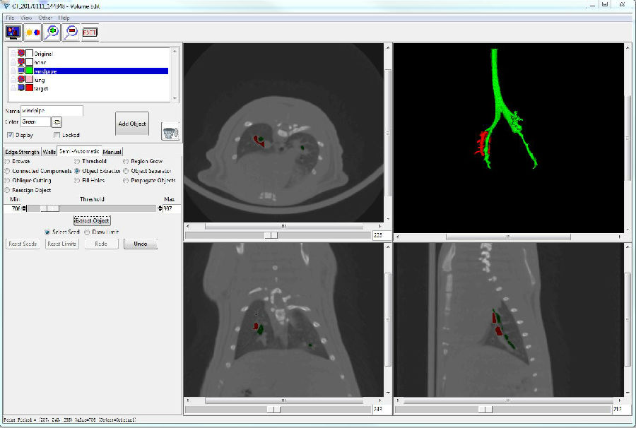
Fig. Three dimensional reconstruction of pulmonary embolism
Customer Providing
Experimental animals or vitro specimens, t
The specific requirements of animal or vitro specimens experiment.


