Catalase Assay Kit
Instruction manual
First Edition (Revised on April, 2016)
[ INTENDED USE ]
The kit is a visible light method for the in vitro quantitative measurement of catalase (CAT) activity in serum, plasma, hemolysate, tissue homogenates, etc..
[ REAGENTS AND MATERIALS PROVIDED ]
Reagents | Quantity(100T-96S) | Reagents | Quantity(100T-96S) |
Reagent 1 | 1×100ml | Reagent 3 | 1 |
Reagent 2 | 1×10ml | Reagent 4 | 1×10ml |
Instruction manual | 1 |
Note: R4 freezes in cold days, when use, please place it in hot water bath until it becomes limpid completely.
[ MATERIALS REQUIRED BUT NOT SUPPLIED ]
1. A spectrophotometer capable of measuring absorbance at 405nm, glass cuvettes of 12.5px light path.
2. Thermostatic water bath or air bath capable of controlling temperature at 37oC.
3. Desk centrifuge.
4. Transferpettor and tips.
5. Vortex mixer.
6. A source of pure water (preferably double distilled water and double distilled water).
[ STORAGE OF THE KITS ]
This kit can be stored at 2-8oC hermetically for 6 months.
Reagent 1: Solution, can be stored at 4oC for 6 months.
Reagent 2: Substrate solution, can be stored at 4oC for 6 months.
Reagent 3: Chromogenic agent powder, can be stored at 4oC for 6 months.
Reagent 4: Solution, can be stored at 4oC for 6 months. It freezes in cold days, when use, please place it in hot water bath until it becomes limpid completely.
[ REAGENT PREPARATION ]
R3 Preparation: Add distilled water until volume reaches to 100ml, dissolve sufficiently. This preparation should be down 1 hour before assay. If there is undissolved powder at bottom, then please take supernant to use directly, this situation will not disturb results.
[ SAMPLE PREPARATION ]
1. Serum
Use a serum separator tube and allow samples to clot for two hours at room temperature or overnight at 4oC before centrifugation for 10 minutes at approximately 2,000rpm. Assay freshly prepared serum immediately or store samples in aliquot at -20oC or -80oC for later use. Avoid repeated freeze/thaw cycles.
2. Plasma
Collect plasma using EDTA or heparin as an anticoagulant. Centrifuge samples for 10 minutes at 2,000rpm at 2-8oC within 30 minutes of collection. Remove plasma and assay immediately or store samples in aliquot at -20oC or -80oC for later use. Avoid repeated freeze/thaw cycles.
3. Hemolysate
Take 50µl whole blood, add distilled water until volume reaches to 5ml, place for 10 minutes and then you can start assay.
4. Tissue homogenates
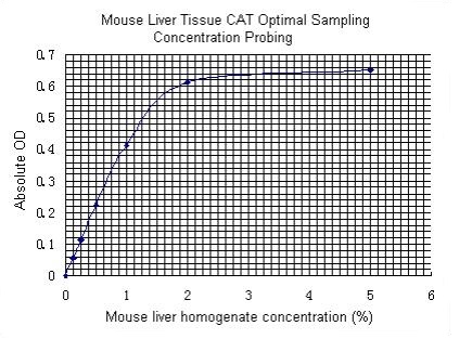
Weigh tissue accurately, add 9 times distilled water to make 10% tissue homogenate, centrifugate at 2500rpm for 10 minutes. Take supernatant, dilute with physiological saline to optimal sampling concentration for assay (you can read optimal sampling concentration probing in appendix).
[ ASSAY PROCEDURE ]
1. Serum/plasma CAT assay
Operation table:
Contrast tube (ml) | Sample tube (ml) | |
Serum/plasma | 0.1 | |
Reagent 1 (37oC prewarmed) | 1.0 | 1.0 |
Reagent 2 (37oC prewarmed) | 0.1 | 0.1 |
Mix sufficiently, place in 37oC water bath for 1 minute accurately. | ||
Reagent 3 | 1.0 | 1.0 |
Reagent 4 | 0.1 | 0.1 |
Serum/plasma | 0.1 | |
Mix sufficiently, place for 10 minutes, transfer in cuvettes of 0.5cm light path, measure OD values of all tubes at 405nm (adjust zero by distilled water). | ||
In order to operate expediently, you can do all preparations at first, label all test tubes by number, add 0.1ml sample and 1ml Reagent 1, then put all test tubes in 37oC water bath for 3~5 minutes. During operation, add 0.1ml Reagent 2, count time accurately at the same time, mix sufficiently immediately, place in 37oC water bath, when t=1min, add chromogenic agent immediately and terminate reaction, mix sufficiently. Then you can operate Tube 2, Tube 3…contrast tube & sample tube. Contrast tube and sample tube must be measured at same time.
2. 1:99 hemolysate CAT assay:
Operation table:
Blank contrast tube (ml) | Sample tube (ml) | Self contrast tube (ml) | |
Distilled water (ml) | 0.05 | 2.1 | |
1:99 hemolysate | 0.05 | 0.05 | |
Reagent 1 (37oC prewarmed) | 1.0 | 1.0 | |
Reagent 2 (37oC prewarmed) | 0.1 | 0.1 | |
| Mix sufficiently, place in 37oC water bath for 1 minute accurately. | |||
Reagent 3 (ml) | 1.0 | 1.0 | |
Mix sufficiently, place for 10 minutes, transfer in cuvettes of 0.5cm light path, measure OD values of all tubes at 405nm (adjust zero by distilled water). | |||
3. Tissue homogenate CAT assay
Operation table:
Contrast tube (ml) | Sample tube (ml) | |
Tissue homogenate | 0.05 | |
Reagent 1 (37oC prewarmed) | 1.0 | 1.0 |
Reagent 2 (37oC prewarmed) | 0.1 | 0.1 |
Mix sufficiently, place in 37oC water bath for 1 minute accurately. | ||
Reagent 3 | 1.0 | 1.0 |
Reagent 4 | 0.1 | 0.1 |
Tissue homogenate | 0.05 | |
Mix sufficiently, place for 10 minutes, transfer in cuvettes of 0.5cm light path, measure OD values of all tubes at 405nm (adjust zero by distilled water). | ||
[ TEST PRINCIPLE ]
Ammonium molybdate can pause H2O2 decomposing reaction catalyzed by catalase (CAT) immediately, residual H2O2 can react with ammonium molybdate to produce a yellowish complex. CAT activity can be calculated by measuring OD value at 405nm.
[ CALCULATION OF RESULTS ]
1. Serum/plasma
a. Definition: 1µmol H2O2 decomposing per ml serum (or plasma) per second is considered as 1 activity unit (U).
b. Formula:

Note: * 271 is reciprocal value of slope.
c. Example: Take 0.1ml serum to measure CAT activity, in results, ODContrast is 0.720, ODSample is 0.675. Calculate as follows:


2. Hemoglobin
a. Definition: 1µmol H2O2 decomposing per mg hemoglobin per second is considered as 1 activity unit (U).
b. Formula:


Note: * 271 is reciprocal value of slope.
c. Example:Take 0.05ml 1:99 mouse hemolysate to measure CAT activity, in results, ODBlank contrast is 0.569, ODSelf contrast is 0.105, ODSample is 0.516, hemoglobin content in 1:99 hemolysate is 1.324mgHb/ml. Calculate as follows:


3. Tissue homogenate:
a. Definition: 1µmol H2O2 decomposing per mg tissue protein per second is considered as 1 activity unit (U).
b. Formula:

Note: * 271 is reciprocal value of slope
c. Example:
① Take 0.05ml 0.5% rat liver tissue homogenate to measure CAT activity, in results, ODContrast is 0.671, ODSample is 0.445, protein content in 0.5% rat liver tissue homogenate is 0.48mgprot/ml Calculate as follows:

② Take 0.05ml 5% earthworm tissue homogenate to measure CAT activity, in results, ODContrast is 0.605, ODSample is 0.425, protein content in 5% earthworm tissue homogenate is 3.1158mgprot/ml. Calculate as follows:

③ Take 0.05ml 10% rice leaf homogenate to measure CAT activity, in results, ODContrast is 0.704, ODSample is 0.332, protein content in 10% rice leaf homogenate is 3.1303mgprot/ml Calculate as follows:

④ Take 0.05ml 0.5% Acipenser sinensis liver tissue homogenate to measure CAT activity, in results, ODContrast is 0.580, ODSample is 0.426, protein content in 0.5% Acipenser sinensis liver tissue homogenate is 0.3063mgprot/ml Calculate as follows:

[ IMPORTANT NOTE ]
1. When you do serum/ plasma CAT assay, if there is no hemolysis in samples, then you just need to take 2 random samples for contrast per batch or use double distilled water for contrast; if there is hemolysis, then you must make contrast tube for each sample.
2. When do tissue homogenate CAT assay, if there is no hyperlipemia, then you just need to take 2 random samples for contrast per batch or use double distilled water for contrast; if there is hyperlipemia, then you must make contrast tube for each sample.
3. When you do whole blood CAT assay, you must make contrast tube for each sample.
[ APPENDIX ]
Optimal sampling volumes:
1. Reagents & preparation: Offered and prepared according to manual by Cloud-Clone Corp.
2. Sample source: Normal mouse liver tissue, make 10% homogenate, then dilute with physiological saline to different concentrations.
3. Operation table:
Contrast tube (ml) | Sample tube (ml) | |
Tissue homogenate | 0.05 | |
Reagent 1 (37oC prewarmed) | 1.0 | 1.0 |
Reagent 2 (37oC prewarmed) | 0.1 | 0.1 |
Mix sufficiently, place in 37oC water bath for 1 minute accurately. | ||
Reagent 3 | 1.0 | 1.0 |
Reagent 4 | 0.1 | 0.1 |
Tissue homogenate | 0.05 | |
Mix sufficiently, place for 10 minutes, transfer in cuvettes of 0.5cm light path, measure OD values of all tubes at 405nm (adjust zero by distilled water). | ||
4. Results
Homogenate concentration(%) | OD1 | OD2 | ODAverage | ΔOD |
0.125 | 0.613 | 0.612 | 0.613 | 0.058 |
0.25 | 0.556 | 0.560 | 0.558 | 0.113 |
0.5 | 0.440 | 0.449 | 0.445 | 0.226 |
1 | 0.258 | 0.258 | 0.258 | 0.413 |
2 | 0.258 | 0.059 | 0.059 | 0.612 |
5 | 0.021 | 0.018 | 0.020 | 0.651 |
10 | 0.032 | 0.031 | 0.032 | 0.639 |
Contrast tube | 0.669 | 0.673 | 0.671 |
5.Mouse Liver tissue optimal sampling concentration probing curve:Referenced sampling concentration is from 0.25% to 1%. In this range, enzyme curve appears direct proportion relatively after regression curve treatment. If sampling concentration is too high or too low, then results won't appear significant deviation after statistical treatment.


