Sample Preparation of Transforming Growth Factor Beta (TGFβ)
Transforming growth factor beta (TGF-β) is a group of transforming growth factor beta superfamily of cytokines, which can control proliferation, cellular differentiation, and other functions in most cells. It is widely located in normal cells and transformed cells. It is a type of cytokine which plays a role in promotion of embryonic development, immunomodulatory effect, growth suppression, wound healing, angiogenesis, extracellular matrix precipitation and so on. This cytokine can promote the transformation of normal fibroblasts from adherent growth to suspension. Therefore, it is named.
TGF-β exists in Mammals at least three isoforms called TGF-β1, TGF-β2 and TGF-β3. The genes of TGF-βs are located in different chromosomes and the amino acide homologies among them range from 70-80%. Among these TGF-βs, the proportion of TGF-β1 is more than 90%. And TGF-β1 is secreted by many cell types including macrophages, lymphocytes, endothelial cells and chondrocytes. It also can be secreted by leukemia cells, mainly distributed in human platelets and mammal bones. While TGF-β2 is secreted by Keratinocytes, granule cell layer and neuroglioma cells. Moreover, TGF-β3 is derived from Mesenchymal cell.
TGF-βs are secreted by many cell types. All three TGF-βs are synthesized as disulfide-linked homodimers containing a propeptide region. After it is synthesized, the TGF-β homodimer interacts with a Latency Associated Peptide (LAP, an N-terminal signal peptide of 20-30 amino acids derived from the TGF beta gene product) forming a complex called Small Latent Complex (SLC). This complex remains in the cell until it is bound by another protein called Latent TGF-β-Binding Protein (LTBP), forming a larger complex called Large Latent Complex (LLC). It is LLC that get secreted to the extracellular matrix (ECM). That is to say, most TFG-β is secreted as a latent form(LLC) in biological samples. The sample is needed to be further processed in order to release TGF-β from LLC. After preparation, the TGF-β level in samples can be accurately measured since the antibodies in the kits is specifically against to the free TGF-β. The sample preparation procedure is listed below.
[ SAMPLE Preparation PROCEDURE ]
Serum/Plasma
1. To 50μL of serum/plasma, add 10μL of 1 N HCl. Mix well.
2. Incubate 10 minutes at room temperature.
3. Neutralize the acidified sample by adding 10μL of 1.2 N NaOH/0.5 M HEPES. Mix well. Add 80μL Standard Diluent. Mix well. Assay immediately.
4. The concentration read off the standard curve must be multiplied by the appropriate dilution factor, 3.
Cell culture supernates
1. To 100μL of cell culture supernate, add 20μL of 1 N HCI. Mix well.
2. Incubate 10 minutes at room temperature.
3. Neutralize the acidified sample by adding 20μL of 1.2 N NaOH/0.5 M HEPES. Mix well. Assay immediately.
4. The concentration read off the standard curve must be multiplied by the dilution factor, 1.4.
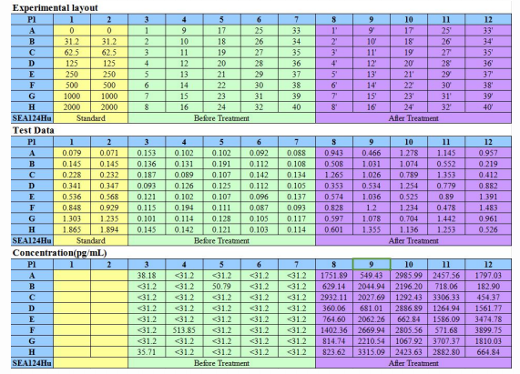
Take ELISA Kit for TGF-β1 (SEA124Hu) as a example, we used it to measure the TGF-β1 levels in samples before and after treatment and made a comparison. As shown in figure 1, the TGF-β1 levels in serum samples after treatment are increased. It is because that the TGF-β1 is released from LLC(the complex of TGF-β1, LAP and LTBP) after treatment. The data may be not precise by directly measure the TGF-β1 levels in samples without treatment. In order to obtain the accurate quantitative measurement of TGF-β1, pre-treatment in samples is recommended before test.

Figure1 The comparison of TGF-β1 level in samples: before and after treatment
The references related to the products of TGF-βs are shown in Table1. Your attention is invited.
Table 1 References of ELISA Kits for TGF-βs Detection
For more information, please visit http://www.cloud-clone.us/.
