A study on establishment of tumor model by subcutaneous injection and intrahepatic injection of Hep G2 cells in nude mice
Primary liver cancer is one of the most common malignant tumors, according to statistics, the annual mortality of liver cancer is second highest of all cancers’ mortality, morbidity and mortality of liver cancer in China is the largest in the world, accounting for 55% of the new incidence and deaths each year. And the number of liver cancer patients is increasing year by year. Because of its high malignant degree, aggressiveness, rapid growth, easy recurrence, metastasis and other biological characteristics, it is known as the 'king of cancer'. Despite the current various surgical, chemotherapy and other treatment methods continue to improve, the effect is still poor. In recent years, with the development of molecular biology and genomics, gene therapy has become the focus of cancer therapy.
The establishment of tumor animal models is an important method to study the pathogenesis of tumor and to carry out tumor treatment. Animal models should be as close as possible to the natural biological processes of human tumors, including tumor genesis, development, invasion, and metastasis. The establishment of a good animal model of liver cancer is a good substitute for the study of the formation of human hepatocellular carcinoma and the anti-tumor immune response.
At present, the transplanted liver cancer model is commonly used in the laboratory, and the transplanted model can be divided into In situ tumor and Ectopic tumor according to the location of the transplanted tumor. By injecting liver cancer cells into nude mice to establish liver orthotopic tumor model, by observing the process and status of In situ tumor formation to simulate the growth of human liver cancer better, and to provide experimental animal model for anti-tumor immunity research. The Ectopic tumor can be formed by subcutaneous injection of liver cancer cells. The operation of subcutaneous tumor is simple, easy to be controlled, has a high success rate, and the volume change of tumor is easy to be measured, and the tumor is easy to be given local intervention.
Cloud-Clone Corp. can provide nude mice In situ tumor and subcutaneous tumor model to you, and according to your specific requirements to complete the experiments.
The experimental method of nude mice In situ tumor formation:
1) HepG2-luc is digested and centrifuged, and approximately 5^106 cells are obtained, then these cells are dissolved in 0.1 mL DMEM culture medium.
2) Use 3% pentobarbital anesthetized nude mice (SPF Balb/C nude mice, male, 4~5 weeks, 15g~20g), expose the liver by opening the abdomen, inject liver cancer cells into nude mice's liver, stitch wounds and place the nude mice in a heating pad, feed them after they were awake.
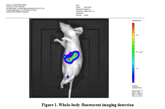
3) Whole-body fluorescent imaging detection. Figure 1 shows the fluorescence.

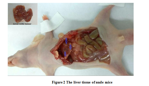
4) Kill nude mice after four weeks, observe the growth of liver tumor. All suspicious organizations are marked and photographed. Figure 2 shows the liver of nude mice.

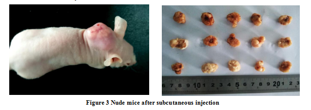
The subcutaneous tumor formation in nude mice is relatively simple. HepG2 cells is digested and centrifuged, and approximately 5^106 cells are gotten, these cells are dissolved in 0.1 mL DMEM culture medium. Use 3% pentobarbital anesthetized nude mice (SPF Balb/C nude mice, male, 4~5 weeks, 15g~20g), liver cancer cells were injected under the skin of the nude mice. About one week after inoculation, the mass began to appear under the skin, and subcutaneous tumor was appeared around 4 weeks. Figure 3 shows nude mice after subcutaneous injection.

For more relevant products, please visit www.cloud-clone.com/.
