An accomplice of the coronavirus-ACE2
Why ACE2 (Angiotensin I Converting Enzyme 2) is called "accomplice" to coronavirus? Let's start with the coronavirus.
The characteristic appearance of virions looks like solar corona or crown by electron microscopy, hence the name coronavirus. The viruses can be divided into four genera (α, β, γ and δ), SARS-CoV, MERS-CoV and SARS-CoV-2 (previously known as 2019-nCoV), belong to the β genus. Figure 1 shows the illustration of the morphology of SARS-CoV-2.
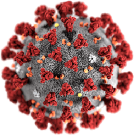
Figure 1. Illustration of the morphology of SARS-CoV-2 (This image is from Wikipedia)
The coronavirus is the largest known RNA virus with the largest genome. Proteins that contribute to the overall structure of coronaviruses are the spike (S), envelope (E), membrane (M), and nucleocapsid (N). Viruses invade the body mainly by binding to receptors on the surface of host cells through S proteins. One of the patterns of invasion is from the respiratory tract into the lungs. There are types I and II alveolar epithelial cells, vascular endothelial cells, macrophages and other immune cells in the alveoli. In theory, any type of cell could be the target cell for coronavirus.
With the preliminary understanding of coronaviruses, you must wonder how does coronavirus relate to ACE2?
In normal lung tissues, ACE2 was found in both type I and type II alveolar epithelial cells, and 80% of ACE2 receptors were mainly clustered in type II alveolar epithelial cells. As mentioned above, S protein invades cells by binding to receptors on the cell surface. Theoretically, without the receptor, the virus cannot invade the cell. Both the well-known SARS-CoV and the more recent SARS-CoV-2 bind to ACE2 through the S protein and invade the lungs. This is the first evidence of ACE2 as an "accomplice".
Besides, some experts believe that the intensity of SARS-CoV-2 infection is related to the Renin-Angiotensin System (RAS). RAS regulation pathways are roughly as follows: Renin carries out the conversion of angiotensinogen to angiotensin I (AngI), then AngI is converted to angiotensin II (AngII) by the angiotensin-converting enzyme (ACE). After that, produced AngII can bind to Angiotensin II Receptor 1 (AT1), thereby increasing pulmonary vascular permeability, promoting proliferation of lung fibroblasts, and inducing apoptosis of alveolar epithelial cells, etc. ACE2 can catalyse the conversion of AngI to Ang 1-9, or the conversion of AngII to Ang 1-7. So the levels of AngI and AngII are reduced. Figure 2 shows the RAS regulatory pathway. According to current data, the most severe and fatal cases of SARS-CoV-2 infection are elderly patients with cardiovascular diseases (including hypertension). Based on RAS regulatory pathway and current data, ACE2 level will be down-regulated after coronavirus infection, resulting in increased AngII level, and then over stimulation of AT1, resulting in increased pulmonary capillary permeability, then resulting in pulmonary edema, acute severe lung injury and acute lung failure, etc. Thus it can be seen that ACE2 can not only help the virus invade cells, but also lead to aggravation of the disease of infected people, which is the second evidence of ACE2 as an "accomplice".
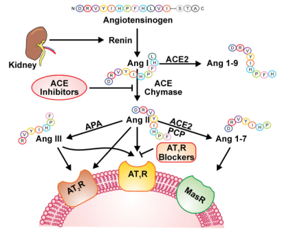
Figure 2. RAS regulatory pathway (Role of the ACE2/Angiotensin 1–7 Axis of the Renin–Angiotensin System in Heart Failure. Circulation Research, 2016 April 15)
Based on the above information, scientists and researchers have come up with several solutions for dealing with or mitigating viral infections. On the one hand, ACE2 protein can be synthesized artificially and then injected into the alveoli through blood vessels, thus interfering with virus recognition and protecting lung cells from being invaded by viruses. On the other hand, ACE2 can be exogenous supplemented to reduce the concentration of AngII, thereby slow the progression of the disease. For example, animal experiments have shown that ACE2 gene knockout mice acute respiratory distress syndrome (ARDS) lesions were significantly worse than the control group, and pulmonary pathology results showed increased vascular permeability and pulmonary edema after ACE2 gene knockout. Moreover, after injecting human recombinant ACE2 protein into ACE2 knockout mice, acute lung injury was improved.
Accordingly, we can know that ACE2 is of great interest to many researchers and doctors who study coronavirus, ARDS, or cardiovascular diseases. For example, the letter "Angiotensin - converting enzymes in acute respiratory distress syndrome" published in Intensive Care Med magazine in March 2019 shows that Department of Intensive Care, Erasme University Hospital, Université Libre de Bruxelles, Brussels, Belgium, chose the ELISA kits for human ACE (SEA004Hu) and ACE2 (SEB886Hu) of Cloud-Clone to detect the changes in the concentration of ACE and ACE2 in plasma, so as to determine the relationship of ARDS with ACE and ACE2 (Figure 3).
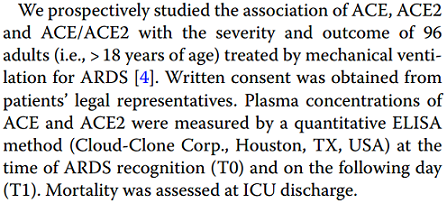
Figure 3. From Intensive Care Med magazine
At present, there are more than 10 citations for ACE2 related to Cloud-Clone products, which indicates that researchers pay attention to ACE2 as well as Cloud-Clone products are recognized by the majority of researchers. Cloud-Clone has a variety of products for targeting ACE2 of different species, including proteins, antibodies and ELISA kits for human, mouse and rat ACE2. The IHC test results (Figure 4-7) and WB test result (Figure 8) of some ACE2 antibodies from Cloud-Clones are shown below.
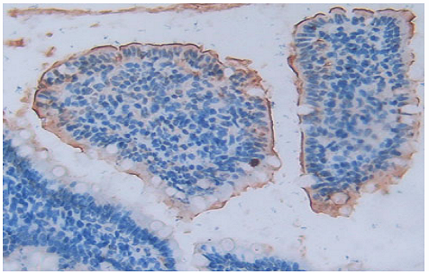
Figure 4. DAB staining on IHC-P; Samples: Rat Small intestine Tissue; Primary Ab: 10µg/ml Rabbit Anti-Rat ACE2 Antibody (Catalog: PAB886Ra01); Second Ab: 2µg/mL HRP-Linked Caprine Anti-Rabbit IgG Polyclonal Antibody (Catalog: SAA544Rb19)
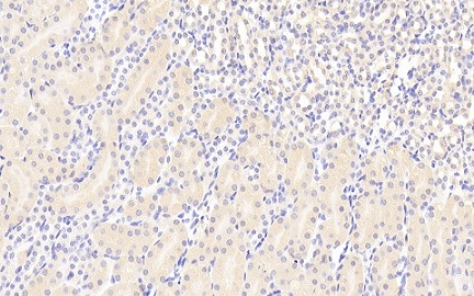
Figure 5. DAB staining on IHC-P; Samples: Mouse Kidney Tissue; Primary Ab: 20µg/ml Rabbit Anti-Mouse ACE2 Antibody (Catalog: PAB886Mu01); Second Ab: 2µg/mL HRP-Linked Caprine Anti-Rabbit IgG Polyclonal Antibody (Catalog: SAA544Rb19)
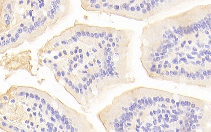
Figure 6. DAB staining on IHC-P; Samples: Mouse Small intestine Tissue; Primary Ab: 20µg/ml Rabbit Anti-Mouse ACE2 Antibody (Catalog: PAB886Mu01); Second Ab: 2µg/mL HRP-Linked Caprine Anti-Rabbit IgG Polyclonal Antibody (Catalog: SAA544Rb19)
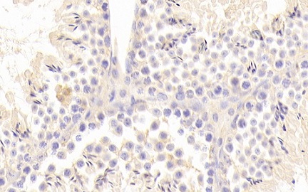
Figure 7. DAB staining on IHC-P; Samples: Mouse Testis Tissue; Primary Ab: 20µg/ml Rabbit Anti-Mouse ACE2 Antibody (Catalog: PAB886Mu01); Second Ab: 2µg/mL HRP-Linked Caprine Anti-Rabbit IgG Polyclonal Antibody (Catalog: SAA544Rb19)
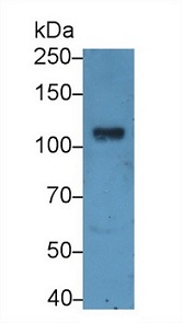
Figure 8. Western Blot; Sample: Porcine Kidney lysate; Primary Ab: 1µg/ml Rabbit Anti-Human ACE2 Antibody (Catalog: PAB886Hu01); Second Ab: 0.2µg/mL HRP-Linked Caprine Anti-Rabbit IgG Polyclonal Antibody (Catalog: SAA544Rb19)
For more information, please visit: http://www.cloud-clone.com/
