Conventional cell proliferation detection techniques
Cells have to split the proliferation of the way. Single-celled organisms, which produce new members by cell division. Multicellular organisms, which produce new cells in a split way to supplement the aging or death cells in the body. Mitosis is the main mode for cell division of eukaryotes. Cell proliferation is the basis of organism growth, development, breeding, genetics, and so on, so cell proliferation detection has always been a hot research topic.
Conventional cell proliferation detection techniques include the following:
(1) Marker protein detection by WB and so on.
PCNA is proliferating cell nuclear antigen, it plays an important role in starting DNA replication and cell proliferation. Ki67 is also a proliferating cell nuclear antigen, it can maintain the stability of the DNA structure in mitosis. It has no expression in the G0 phase of the cell cycle, begins to express in the G1 phase, has gradually increased expression level in the S phase and G2 phase, has the highest expression level in the M phase, and will be degraded after the mitosis. The researchers can know the cell proliferation indirectly by detecting the expression of PCNA or Ki67 by WB.
Advantages: simplicity, universality, tissue non-specificity.
Disadvantage: the state of the cell proliferation is showed indirectly.
(2) EdU detection
EdU (5-Ethynyl-2'-deoxyuridine) is a thymidine analogue which can be incorporated into the DNA of dividing cells. The DNA replication activity can be known as the EdU is detected with a fluorescent dyes. BrdU (bromodeoxyuridine) detection is largely replaced by EdU detection now.
Advantages: in vitro test, high detection sensitivity, other labelled antibodies or fluorescent proteins can be detected at the same time.
Disadvantage: toxicity.
(3) CFSE detection
CFSE (carboxyfluorescein succinimidyl amino ester) is fluorescent dye, it can penetrate the cell membrane and enter the cell, irreversibly combine with amino and be coupled to the protein. When cells divide, fluorescent is divided equally to two daughter cells, the fluorescence intensity of daughter cells is half of parental cells’. The fluorescent of CFSE can be excited by flow cytometry 488 nm exciting light.
Advantages: the direct method, living cell detection.
Disadvantages: when the concentration of CFSE is too low, the fluorescence intensity is not enough to be detected; when its concentration is too high, the toxicity will affect cell proliferation.
(4) Cell Counting Kit-8 detection
Cell Counting Kit-8 (CCK8) detection is upgrade version of MTT assay. WST-8 (2-(2-methoxy-4-nitrophenyl)-3-(4-nitrophenyl)-5-(2,4-disulfophenyl)-2H-tetrazolium) is the CCK8 solution, and it is similar with MTT, can be reduced by dehydrogenase which is in the mitochondria. However, the CCK8 assay yield a water-soluble formazan. The faster and more the cell proliferation, the darker the color. Run the microplate reader and conduct measurement at 450nm.
Advantages: simplicity, intuitive result, living cell detection.
Disadvantages: the number of living cell is gotten indirectly. CCK8 solution color is light red, the color is closed to the color of culture medium with phenol red, so it is easy to add more or forget to add.
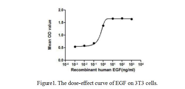
Cloud-Clone Corp. has pushed out active proteins, the CCK8 detection in combination with microscopy observation were used for the detection of effect of active protein on cell proliferation. Take active epidermal growth factor (APA560Hu02) for example, when its concentration increases, it can stimulate the 3T3 cell proliferation. The result is shown below.

For more products and information, please visit: http://www.cloud-clone.com/.
