New findings on potential treatments for Parkinson's disease
Parkinson's disease (PD) is the second most common progressive neurodegenerative disease and its incidence is expected to increase as the global population ages. Resulting from a pathophysiologic loss or degeneration of dopaminergic neurons in the substantia nigra of the midbrain and the development of neuronal Lewy Bodies, idiopathic PD is associated with risk factors including aging, family history, pesticide exposure and environmental chemicals. Given that aggregated and misfolded a-synuclein (a-Syn) species are the major constituents of Lewy bodies, PD is classified as a synucleinopathy. Numerous therapies have been developed that provide meaningful symptomatic benefits for the classical motor features of the disease. Still, no therapy has been established to slow or stop disease progression. Recently, multiple papers have reported potential treatment methods for PD, which may help further improve the prognosis of PD patients.
1. Co-transplantation of autologous Treg cells in a cell therapy for PD
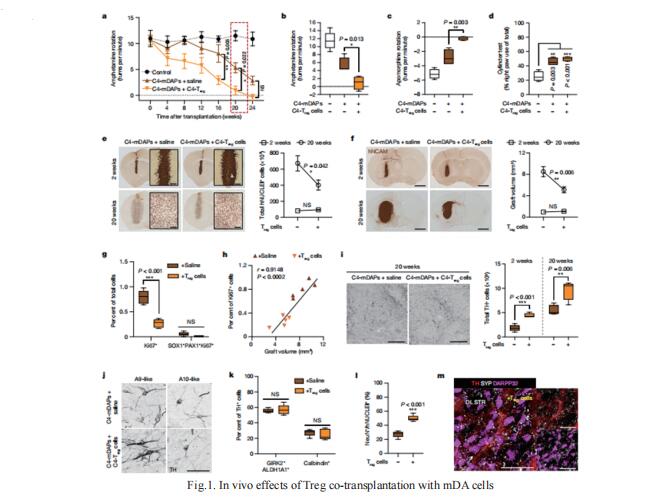
The specific loss of midbrain dopamine neurons (mDANs) causes major motor dysfunction in PD, which makes cell replacement a promising therapeutic approach. However, poor survival of grafted mDANs remains an obstacle to successful clinical outcomes. Kwang-Soo Kim, Molecular Neurobiology Laboratory, Department of Psychiatry and McLean Hospital, Harvard Medical School, USA, and his team showed that the surgical procedure itself (referred to here as ‘needle trauma’) triggers a profound host response that is characterized by acute neuroinflammation, robust infiltration of peripheral immune cells and brain cell death[1]. When midbrain dopamine (mDA) cells derived from human induced pluripotent stem (iPS) cells were transplanted into the rodent striatum, less than 10% of implanted tyrosine hydroxylase (TH)+ mDANs survived at two weeks after transplantation. By contrast, TH- grafted cells mostly survived. Notably, transplantation of autologous regulatory T (Treg) cells greatly modified the response to needle trauma, suppressing acute neuroinflammation and immune cell infiltration. Furthermore, intra-striatal co-transplantation of Treg cells and human-iPS-cell-derived mDA cells significantly protected grafted mDANs from needle-trauma-associated death and improved therapeutic outcomes in rodent models of PD with 6-hydroxydopamine lesions (Fig.1). Together, these data emphasize the importance of the initial inflammatory response to surgical injury in the diferential survival of cellular components of the graft, and suggest that co-transplanting autologous Treg cells effectively reduces the needle-trauma-induced death of mDANs, providing a potential strategy to achieve better clinical outcomes for cell therapy in PD.

2. Development of an a-Syn positron emission tomography tracer for imaging synucleinopathies
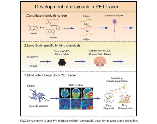
Synucleinopathies are characterized by the accumulation of a-Syn aggregates in the brain. Positron emission tomography (PET) imaging of synucleinopathies requires radiopharmaceuticals that selectively bind a-Syn deposits. Keqiang Ye, Department of Pathology and Laboratory Medicine, Emory University School of Medicine, USA, and his team reported the identification of a brain permeable and rapid washout PET tracer [18F]-F0502B, which showed high binding affinity for a-Syn, but not for Ab or Tau fibrils, and preferential binding to a-Syn aggregates in the brain sections[2]. Employing several cycles of counter screenings with in vitro fibrils, intraneuronal aggregates, and neurodegenerative disease brain sections from several mice models and human subjects, [18F]-F0502B images a-Syn deposits in the brains of mouse and non-human primate PD models. They further determined the atomic structure of the a-Syn fibril-F0502B complex by cryo-EM and revealed parallel diagonal stacking of F0502B on the fibril surface through an intense noncovalent bonding network via inter-ligand interactions(Fig.2). Therefore, the study reveals a promising lead compound for imaging a-Syn and facilitating the diagnosis of synucleinopathies. This might provide a tool to improve the understanding of disease progression and potentially monitor therapeutic efficacy in clinical trials.

3. The c-Abl inhibitor IkT-148009 suppresses neurodegeneration in mouse models of heritable and sporadic PD
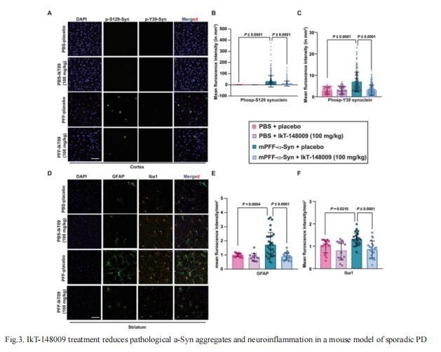
Animal models of PD suggest that activation of Abelson tyrosine kinase (c-Abl) plays an essential role in the initiation and progression of a-Syn pathology and initiates processes leading to degeneration of dopaminergic and nondopaminergic neurons. Given the potential role of c-Abl in PD, Valina L. Dawson, Neuroregeneration and Stem Cell Programs, Institute for Cell Engineering, Johns Hopkins University School of Medicine, USA, and his team developed a c-Abl inhibitor library to identify orally bioavailable c-Abl inhibitors capable of crossing the blood-brain barrier based on predefined characteristics, leading to the discovery of IkT-148009[3]. In mouse models of both inherited and sporadic PD, IkT-148009 suppressed c-Abl activation to baseline and substantially protected dopaminergic neurons from degeneration when administered therapeutically by once daily oral gavage beginning 4 weeks after disease initiation. Recovery of motor function in PD mice occurred within 8 weeks of initiating treatment concomitantly with a reduction in a-Syn pathology in the mouse brain (Fig.3). These findings suggest that IkT-148009 may have potential as a disease-modifying therapy in PD.

References
[1]Park TY, Jeon J, Lee N, et al. Co-transplantation of autologous Treg cells in a cell therapy for Parkinson's disease. Nature. 2023;619(7970):606-615. (IF=64.8)
[2]Xiang J, Tao Y, Xia Y, et al. Development of an α-synuclein positron emission tomography tracer for imaging synucleinopathies. Cell. 2023;S0092-8674(23)00644-X. (IF=64.5)
[3]Karuppagounder SS, Wang H, Kelly T, et al. The c-Abl inhibitor IkT-148009 suppresses neurodegeneration in mouse models of heritable and sporadic Parkinson's disease. Sci Transl Med. 2023;15(679):eabp9352. (IF=17.1)
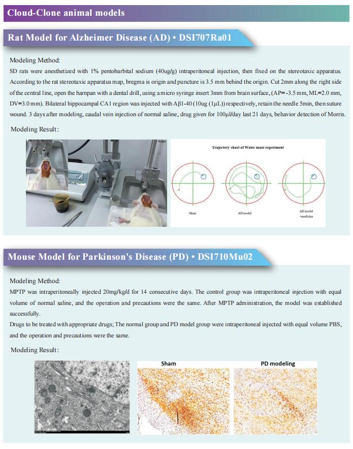
Cloud-Clone can not only provide animal models of various neurological diseases, including Parkinson's disease, Alzheimer's disease, anxiety disorder, chronic stress depression, etc., cover common neurological diseases. We also have various neurological disease detection indicators related products, which can help the majority of scientific researchers to carry out neurological disease related research.

