Packages (Simulation)

Reagent Preparation

Image (I)
Image (II)
Certificate


Mini Samples ELISA Kit for Vascular Endothelial Growth Factor C (VEGFC)
VEGF-C; Flt4-L; VRPL; Vascular Endothelial Growth Factor-Related Protein
- Product No.MEA145Hu
- Organism SpeciesHomo sapiens (Human) Same name, Different species.
- Sample TypeSerum, plasma, tissue homogenates, cell lysates, cell culture supernates and other biological fluids
- Test MethodDouble-antibody Sandwich
- Assay Length3h
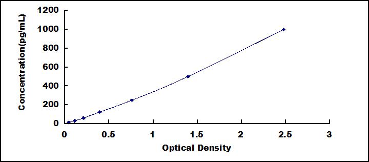
- Detection Range15.6-1,000pg/mL
- SensitivityThe minimum detectable dose of this kit is typically less than 5.5pg/mL.
- DownloadInstruction Manual
- UOM 48T96T 96T*5 96T*10 96T*100
- FOB
US$ 419
US$ 598
US$ 2691
US$ 5083
US$ 41860
For more details, please contact local distributors!
Specificity
This assay has high sensitivity and excellent specificity for detection of Mini Samples Vascular Endothelial Growth Factor C (VEGFC).
No significant cross-reactivity or interference between Mini Samples Vascular Endothelial Growth Factor C (VEGFC) and analogues was observed.
Recovery
Matrices listed below were spiked with certain level of recombinant Mini Samples Vascular Endothelial Growth Factor C (VEGFC) and the recovery rates were calculated by comparing the measured value to the expected amount of Mini Samples Vascular Endothelial Growth Factor C (VEGFC) in samples.
| Matrix | Recovery range (%) | Average(%) |
| serum(n=5) | 89-104 | 94 |
| EDTA plasma(n=5) | 88-97 | 94 |
| heparin plasma(n=5) | 81-95 | 84 |
Precision
Intra-assay Precision (Precision within an assay): 3 samples with low, middle and high level Mini Samples Vascular Endothelial Growth Factor C (VEGFC) were tested 20 times on one plate, respectively.
Inter-assay Precision (Precision between assays): 3 samples with low, middle and high level Mini Samples Vascular Endothelial Growth Factor C (VEGFC) were tested on 3 different plates, 8 replicates in each plate.
CV(%) = SD/meanX100
Intra-Assay: CV<10%
Inter-Assay: CV<12%
Linearity
The linearity of the kit was assayed by testing samples spiked with appropriate concentration of Mini Samples Vascular Endothelial Growth Factor C (VEGFC) and their serial dilutions. The results were demonstrated by the percentage of calculated concentration to the expected.
| Sample | 1:2 | 1:4 | 1:8 | 1:16 |
| serum(n=5) | 79-104% | 79-93% | 90-103% | 96-105% |
| EDTA plasma(n=5) | 90-104% | 87-101% | 86-94% | 90-98% |
| heparin plasma(n=5) | 92-104% | 78-90% | 81-94% | 81-88% |
Stability
The stability of kit is determined by the loss rate of activity. The loss rate of this kit is less than 5% within the expiration date under appropriate storage condition.
To minimize extra influence on the performance, operation procedures and lab conditions, especially room temperature, air humidity, incubator temperature should be strictly controlled. It is also strongly suggested that the whole assay is performed by the same operator from the beginning to the end.
Reagents and materials provided
| Reagents | Quantity | Reagents | Quantity |
| Pre-coated, ready to use 96-well strip plate | 1 | Plate sealer for 96 wells | 4 |
| Standard | 2 | Standard Diluent | 1×20mL |
| Detection Reagent A | 1×60µL | Assay Diluent A | 1×6mL |
| Detection Reagent B | 1×60µL | Assay Diluent B | 1×6mL |
| TMB Substrate | 1×4.5mL | Stop Solution | 1×3mL |
| Wash Buffer (30 × concentrate) | 1×10mL | Instruction manual | 1 |
Assay procedure summary
1. Prepare all reagents, samples and standards;
2. Add 25µL standard or sample to each well. Incubate 1 hour at 37°C;
3. Aspirate and add 25µL prepared Detection Reagent A. Incubate 1 hour at 37°C;
4. Aspirate and wash 3 times;
5. Add 25µL prepared Detection Reagent B. Incubate 30 minutes at 37°C;
6. Aspirate and wash 5 times;
7. Add 25µL Substrate Solution. Incubate 10-20 minutes at 37°C;
8. Add 20µL Stop Solution. Read at 450nm immediately.
GIVEAWAYS
INCREMENT SERVICES
| Magazine | Citations |
| Journal of Cellular Biochemistry | Alternatively activated RAW264. 7 macrophages enhance tumor lymphangiogenesis in mouse lung adenocarcinoma PubMed: 19241443 |
| genetics and molecular research | Expression of COX-2 and VEGF-C in cholangiocarcinomas at different clinical and pathological stages PubMed: 26125824 |
| Experimental and Therapeutic | Fenofibrate inhibits the expression of VEGFC and VEGFR-3 in retinal pigmental epithelial cells exposed to hypoxia PubMed: 26622498 |
| Am J Pathol | IL-10 Indirectly Regulates Corneal Lymphangiogenesis and Resolution of Inflammation via Macrophages PubMed: 26608451 |
| PLoS One. | Interleukin-6 Induces Vascular Endothelial Growth Factor-C Expression via Src-FAK-STAT3 Signaling in Lymphatic Endothelial Cells. pubmed:27383632 |
| Oncology Letters | RNAi-mediated gene silencing of vascular endothelial growth factor C suppresses growth andinduces apoptosis in mouse breast cancer in vitro and in vivo. pubmed:27895746 |
| Angiogenesis | Characterization of isolated liver sinusoidal endothelial cells for liver bioengineering Pubmed:29582235 |
| Oncology Reports | Effects of diphyllin as a novel V-ATPase inhibitor on TE-1 and ECA-109 cells Pubmed:29328465 |
| LABORATORY INVESTIGATION | Dynamic signature of lymphangiogenesis during acute kidney injury and chronic kidney disease Pubmed: 31019289 |
| Cutaneous and Ocular Toxicology | Isotretinoin does not alter VEGF-A and VEGF-C levels: Do retinoids behave differently in dose-dependent and/or in vivo/in vitro conditions? Pubmed: 32722957 |


