- SPF
Rat Model for Vascular Calcification (VC)
- Product No.DSI845Ra01
- Organism SpeciesRattus norvegicus (Rat) Same name, Different species.
- Prototype SpeciesHuman
- Sourceinduced by Vitamin D3
- Model Animal StrainsSD Rats(SPF), healthy, male, body weight 180g~200g.
- Modeling Grouping1.Randomly divided into six group: Control group, Model group, Positive drug group and Tested drug group.
- Modeling Period8 weeks
- Modeling MethodDaily vitamin D3 300,000 IU/kg body weight intramuscular injection for 4 weeks.
Nicotine gavage twice a day: Dissolve nicotine in peanut oil and intragastric administration at a dose of 25mg/kg, repeated again after 9 hours for four weeks.Note: Vitamin D3 mainly causes vascular calcification, nicotine stimulates the heartbeat, accelerates the blood pressure, and increases the blood pressure. Animal state should be observed after every intragastric administration. Mice will twitch after intragastric administration, which needs to be alleviated by manual chest compression.
Plasma preparation: At the end of model preparation, rats in each group were anesthetized, and blood was taken from abdominal aorta and injected into test tubes containing 30ul 7.5%EDTA disodium and 40ul aprotinin for centrifugation. Store at -20℃ for testing.
Tissue preparation: The rats were sacrificed after blood was taken, and the thoracic aorta segment of the rats was inserted into 4% paraformaldehyde fixative, dehydrated, embedded in paraffin, and sectioning for use.
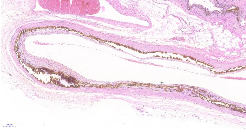
The pathological tissues were stained with Von Kossa. - ApplicationsDisease Model
- Downloadn/a
- UOM Each case
- FOB
US$ 260
For more details, please contact local distributors!
Model Evaluation
1. Determination of vascular calcium content: 10mg of abdominal aorta was dissolved in nitric acid for digestion and drying, and redissolved in deionized water containing 27nmol /LKCl and 27μmol/L LaCl3. Absorbance was read at 442.7nm by atomic spectrophotometer and converted to tissue calcium content (expressed as μmol/gdw).
2. ALP activity determination: Abdominal aorta homogenate was taken, centrifuged at 8000×g for 10 min, supernatant was taken, and protein was quantified by Coomassie bright blue method.ALP activity of vascular tissue was measured according to kit instructions. Absorbance was read at 405nm by UV spectrophotometer. Specific steps: add buffer solution, incubate at 37℃ for 30min, NaOH terminated the reaction, and determine the absorption value at 405nm.ALP activity was calculated using P2 nitrophenol as the standard value, and 1 unit represented the activity of producing 1nM nitrophenol within 30min.Total protein was quantified by BCA method and ALP activity was corrected.
Pathological Results
Von Kossa staining of pathological tissues:
The thoracic aorta of rats was embedded in paraffin and sectionalized. The sections were 4μm thick and dewaxed and dehydrated conventionally.
Soaked in 1% silver nitrate solution and exposed to sunlight for 30 minutes,the slides were immersed in 5% sodium thiosulfate solution for 1min, and then backdyed with basic fuchsin.
Dehydrated, transparent, sealed, observed with light microscope.
Calculation of percentage of aortic calcification area: take the aortic arch to the thoracic aortic segment vessel 1cm, Von Kossa staining after cutting along the longitudinal axis, and Image-pro Plus 6.0 pathological Image analysis software was used to calculate the percentage of calcified plaque area of rats in each experimental group.
RUNX2, BMP2, RANK/RANKL/OPG levels were observed by immunohistochemical method: DAB staining, hematoxylin restaining, conventional gradient ethanol dehydration, neutral gum sealing tablet observation. Negative control was stained with PBS instead of primary antibody.
Three colon sections were randomly taken from each rat, and 5 high power fields were randomly selected from each section under the optical microscope. The ratio of the integral optical density (IOD) value of positive tissue cells in each field to the detection area, namely the average optical density value (MOD), was calculated to represent the staining intensity.
Cytokines Level
Western blot analysis of RUNX2, BMP2, RANK/RANKL/OPG protein expression.
Statistical Analysis
SPSS software is used for statistical analysis, measurement data to mean ± standard deviation (x ±s), using t test and single factor analysis of variance for group comparison, P<0.05 indicates there was a significant difference, P<0.01 indicates there are very significant differences.
GIVEAWAYS
INCREMENT SERVICES
-
 Primary Cells Customized Service
-
 Total Protein/DNA/RNA Extract Customized Service
-
 Tissue/Sections Customized Service
-
 Serums Customized Service
-
 Immunohistochemistry (IHC) Experiment Service
-
 Real Time PCR Experimental Service
-
 Nitric Oxide Assay Kit (A012)
-
 Nitric Oxide Assay Kit (A013-2)
-
 Total Anti-Oxidative Capability Assay Kit(A015-2)
-
 Total Anti-Oxidative Capability Assay Kit (A015-1)
-
 Cloud-Clone Multiplex assay kits
| Catalog No. | Related products for research use of Rattus norvegicus (Rat) Organism species | Applications (RESEARCH USE ONLY!) |
| DSI845Ra01 | Rat Model for Vascular Calcification (VC) | Disease Model |
| TSI845Ra85 | Rat Arterial vessels Tissue of Vascular Calcification (VC) | Paraffin slides for pathologic research: IHC,IF and HE,Masson and other stainings |


