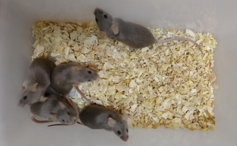
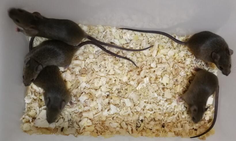
Mouse Model for Spontaneous Abortion (SA)
RSA;Miscarriage; Pregnancy loss;Recurren Spontaneous Abortion
- Product No.DSI638Mu01
- Organism SpeciesMus musculus (Mouse) Same name, Different species.
- Prototype SpeciesHuman
- SourceFemale CBA/J and male DBA/2 for RSA model
- Model Animal StrainsSPF Female CBA/J, male DBA/2, male BALB/c mice
- Modeling GroupingNormal pregnancy control group, healthy pregnancy control group, RSA model group, RSA treatment group.
- Modeling Period6~8 weeks
- Modeling MethodEstablishment of RSA mice model
The normal pregnancy mouse model was established by caging female CBA/J and male BALB/ C mice at a ratio of 2:1, and RSA mice model (recurrent abortion) was established by caging female CBA/J and male DBA/2 mice at a ratio of 2:1. The detection of negative embolus was calculated as day 0 of pregnancy.
The experiment was divided into 4 groups:
Treatment group (female CBA/J, male DBA/2 5)
RSA model group (female CBA/J, male DBA/2 5)
Normal pregnancy control group (female CBA/J 10, male BALB/ C 5)
Healthy non-pregnant control group (female CBA/J 10)
The rats in each group were given intragastric administration from day 0 of gestation, with 10 females in each group.
The dosage was calculated according to the clinical dosage of 70kg adult according to the animal body surface area, and the volume of intragastric administration was 0.4ml /20g. The normal pregnancy group and the RSA model control group were given the same volume of distilled water for 14 days. The mice were sacrificed 1h after the last administration, and their peripheral blood and decidua tissues were collected. - ApplicationsUsed to study the medicine for prevention and treatment of RSA
- Downloadn/a
- UOM Each case
- FOB
US$ 240
For more details, please contact local distributors!
Model Evaluation
The proportion of Th17/Treg cells in PBMCs was detected by flow cytometry:
After PBMCs were isolated, phorbol ester, Ionomycin and Monensin were added at working concentration and incubated at 37°C for 4-6h. The same type of monitoring and detection tubes were set. After Fc receptor blocker was closed, CD4 surface antibody was added for staining, and mixed. After 15min incubation at room temperature, fixation solution and broken membrane/haemolysis were successively added to each tube according to operating requirements. After washing, 2 matching antibodies were added to the experimental tube, and homotype control antibodies were added to the care. After incubation in dark, detergent was added to centrifuge the supernatant, and the cells were re-suspended for testing within 24 hours. CD4 histogram was used to shoot, and Th17 cells and Treg cells were represented by positive scatter plot staining. Cellsquest software was used to obtain and analyze data. (One sample was divided into two staining regimens, Th17: CD4-FITC, IL-17-PE; Treg: CD4-FITC, CD25-APC, FOXP3-PE)
Expression of FOXP3, RORrt, STAT3 and STAT5 in PBMCs detected by RT-PCR:
Total PBMCs RNA was extracted from each group, and RNA purity and integrity were identified. The primers were designed and retrotranscribed into cDNA, using β-actin as internal reference, and then real-time fluorescence quantitative RT-PCR was performed. The relative quantities of FOXP3, RORrt, STAT3 and STAT5 mRNA were calculated by Ct using relative quantitative method.
Pathological Results
1. Histopathological changes of local tissues and cells were observed by HE staining:
Decidua tissues of mice in each group were embedded in paraffin and stained with HE.
2. Observation of apoptosis and pathological changes in local tissues by electron microscopy:
The decidua tissues of mice in each group were taken aseptically, pre-fixed with 2.5% glutaraldehyde, fixed with 1% osmium acid, dehydrated with ethanol and acetone step by step, embedded with resin, and prepared into ultra-thin sections. The decidua tissues of mice in each group were stained with uranium dioxy-lead nitrate solution, and observed under electron microscopy.
3. Immunohistochemical analysis of cytokines STAT3 and STAT5 expression in local tissues:
4. MRNA expressions of FOXP3, RORrt, STAT3 and STAT5 in decidua tissues were detected by RT-PCR.
5. The STAT3 and STAT5 protein expressions in decidua tissues were detected by Western blot.
Cytokines Level
6.1ELISA was used to detect IL-2 and IL-6 in the homogenate of demembraned tissues.
The decidua tissues of each group were collected, and the supernatant was taken for ELISA detection after homogenization.
6.2 Expression of cytokines IL-2, IL-6 and TGF-β1 detected by ELISA:
Serum of each mouse was collected, il-2, IL-6 and TGF-β1 ELISA Kit were used according to the kit instructions. Standard curves were set on the microplate reader, and 3 multiple Wells were set for each specimen and standard product. The absorbance value (A) of serum samples was read by A microplate reader, and the sample concentration value was calculated by standard curve.
Statistical Analysis
SPSS software is used for statistical analysis, measurement data to mean ± standard deviation (x ±s), using t test and single factor analysis of variance for group comparison, P<0.05 indicates there was a significant difference, P<0.01 indicates there are very significant differences.
GIVEAWAYS
INCREMENT SERVICES
-
 Tissue/Sections Customized Service
-
 Serums Customized Service
-
 Immunohistochemistry (IHC) Experiment Service
-
 Small Animal In Vivo Imaging Experiment Service
-
 Small Animal Micro CT Imaging Experiment Service
-
 Small Animal MRI Imaging Experiment Service
-
 Small Animal Ultrasound Imaging Experiment Service
-
 Transmission Electron Microscopy (TEM) Experiment Service
-
 Scanning Electron Microscope (SEM) Experiment Service
-
 Learning and Memory Behavioral Experiment Service
-
 Anxiety and Depression Behavioral Experiment Service
-
 Drug Addiction Behavioral Experiment Service
-
 Pain Behavioral Experiment Service
-
 Neuropsychiatric Disorder Behavioral Experiment Service
-
 Fatigue Behavioral Experiment Service
-
 Nitric Oxide Assay Kit (A012)
-
 Nitric Oxide Assay Kit (A013-2)
-
 Total Anti-Oxidative Capability Assay Kit(A015-2)
-
 Total Anti-Oxidative Capability Assay Kit (A015-1)
-
 Superoxide Dismutase Assay Kit
-
 Fructose Assay Kit (A085)
-
 Citric Acid Assay Kit (A128 )
-
 Catalase Assay Kit
-
 Malondialdehyde Assay Kit
-
 Glutathione S-Transferase Assay Kit
-
 Microscale Reduced Glutathione assay kit
-
 Glutathione Reductase Activity Coefficient Assay Kit
-
 Angiotensin Converting Enzyme Kit
-
 Glutathione Peroxidase (GSH-PX) Assay Kit
-
 Cloud-Clone Multiplex assay kits
| Catalog No. | Related products for research use of Mus musculus (Mouse) Organism species | Applications (RESEARCH USE ONLY!) |
| DSI638Mu01 | Mouse Model for Spontaneous Abortion (SA) | Used to study the medicine for prevention and treatment of RSA |


