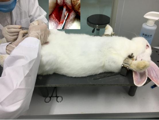
Rabbit Model for Tendon Damage (TD)
Tendon adhesion; Tendon injury
- Product No.DSI794Rb01
- Organism SpeciesOryctolagus cuniculus (Rabbit) Same name, Different species.
- Prototype SpeciesHuman
- SourceInduced by amputation of tendon
- Model Animal StrainsJapanese large white rabbits, healthy, male, age:3 months, weight 2.4~2.6kg.
- Modeling GroupingRandomly divided into six groups: Control group, Model group, Positive drug group and Test drug group(low, medium, high).
- Modeling Period4~6 weeks
- Modeling Method1. Rabbits were anesthetized by intravenous injection of 3% pentobarbital sodium (1ml/kg) via the ear margin.
2. The rabbit right hind legs with shaver shaving, alcohol iodineWineDisinfection, legs stretched fixed on the operating table.
3. In the right foot second toe, fourth toe proximal phalanx linear incision, cut the skin and subcutaneous tissue, layer by layer separation to cut tendon sheath, sheath incision to pick 1 deep flexor tendon and 2 superficial flexor tendon, transection, preparation of transection model.
4. Suture of flexor digitorum profundus tendon, superficial flexor tendon sheath and skin suture without treatment, with iodine disinfection.
5. Postoperative penicillin 400 thousand U/day every rabbit, continuous intramuscular injection for 3 days, to prevent infection.
6. Specimen collection: the subcutaneous tissue or adhesion tissue was cut into the tendon surface by intravenous anesthesia with pentobarbital sodium at 2, 4 and 6 weeks after operation. The left posterior, second toe and fourth toe tendons were taken as the control. - ApplicationsUsed for the prevention and treatment of tendon adhesion
- Downloadn/a
- UOM Each case
- FOB
US$ 300
For more details, please contact local distributors!
Model Evaluation
1. General observation of degree and extent of adhesion of tendon :
adhesion degree and extent of tendon in general specimens and healing condition. And the adhesion score criteria are as follows:
Grade 0: surgical site without adhesion;
Grade 1: a small amount of membranous, band like adhesions confined to sutures where tendon slippage is limited;
Grade 2: small or broad band adhesions limited to sutures where tendon gliding is limited;
Grade 3: large area of adhesion, spread to both ends of the suture point, the tendon is limited or almost impossible to slide;
Grade 4: tendon and sheath tube wall, subcutaneous formation of closely linked, wide range, tendon without any sliding.
Pathological Results
HE staining: the tendon tissue were fixed in 4% paraformaldehyde and embedded in paraffin and cut into slices; stained with HE, observed after the experimental side, under optical microscope and polarizing microscope control side of tendon adhesion histological changes and collagen fibers distribution.
IHC: Stained with Ki67, and observed under the microscope.
Cytokines Level
Statistical Analysis
SPSS software is used for statistical analysis, measurement data to mean ± standard deviation (x ±s), using t test and single factor analysis of variance for group comparison, P<0.05 indicates there was a significant difference, P<0.01 indicates there are very significant differences.
GIVEAWAYS
INCREMENT SERVICES
-
 Tissue/Sections Customized Service
-
 Serums Customized Service
-
 Immunohistochemistry (IHC) Experiment Service
-
 Small Animal In Vivo Imaging Experiment Service
-
 Small Animal Micro CT Imaging Experiment Service
-
 Small Animal MRI Imaging Experiment Service
-
 Small Animal Ultrasound Imaging Experiment Service
-
 Transmission Electron Microscopy (TEM) Experiment Service
-
 Scanning Electron Microscope (SEM) Experiment Service
-
 Learning and Memory Behavioral Experiment Service
-
 Anxiety and Depression Behavioral Experiment Service
-
 Drug Addiction Behavioral Experiment Service
-
 Pain Behavioral Experiment Service
-
 Neuropsychiatric Disorder Behavioral Experiment Service
-
 Fatigue Behavioral Experiment Service
-
 Nitric Oxide Assay Kit (A012)
-
 Nitric Oxide Assay Kit (A013-2)
-
 Total Anti-Oxidative Capability Assay Kit(A015-2)
-
 Total Anti-Oxidative Capability Assay Kit (A015-1)
-
 Superoxide Dismutase Assay Kit
-
 Fructose Assay Kit (A085)
-
 Citric Acid Assay Kit (A128 )
-
 Catalase Assay Kit
-
 Malondialdehyde Assay Kit
-
 Glutathione S-Transferase Assay Kit
-
 Microscale Reduced Glutathione assay kit
-
 Glutathione Reductase Activity Coefficient Assay Kit
-
 Angiotensin Converting Enzyme Kit
-
 Glutathione Peroxidase (GSH-PX) Assay Kit
-
 Cloud-Clone Multiplex assay kits
| Catalog No. | Related products for research use of Oryctolagus cuniculus (Rabbit) Organism species | Applications (RESEARCH USE ONLY!) |
| DSI794Rb01 | Rabbit Model for Tendon Damage (TD) | Used for the prevention and treatment of tendon adhesion |


